
Advanced tumor models shouldn’t require an advanced workflow.
The best 3D models on the market today effectively ask you to become a tissue engineer to use them, a barrier that keeps them out of most pipelines. TissueTinker is built the other way around. It drops into the lab you already run: your well plates, your assays, your microscopy, your culture media, all unchanged. What changes is what you can see.
Three parts, one workflow
The system is three components that work as one.
TTB Bioprinter
A low-cost bioprinter designed to be used, not commissioned. No specialist setup; from box to first print in minutes, inside the culture hood you already have.
TTB Digital Model Library
A software library of ready-to-print tissue geometries, pre-designed for different experimental needs, so you start from a validated model, not a blank build plate.
TTB-Series Bioinks
Tissue-derived matrices that carry the mechanical and biochemical properties of real extracellular matrix. The heart of the system.
The workflow
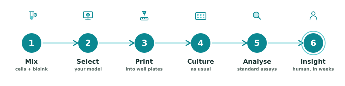
Steps 4 through 6 use the tools you already have. Mix your cells, choose your model, and the printer takes it from there: you culture, analyse, and gain insight exactly as before.
Inside the bioinks
If the system has a heart, it’s the bioink. Generic scaffolds mostly hold cells up against gravity. Ours recreate the environment cells actually respond to.
Biochemical authenticity
Unlike reconstituted collagen, TTB-Series bioinks retain the complex proteomic signature of real tissue, confirmed by mass spectrometry. The cues cells are built to read are actually present:
- Structural proteins: collagens I, III, and IV, in physiological ratios.
- Adhesion molecules: laminin and fibronectin, for integrin-mediated signaling.
- Soluble factors: glycosaminoglycans that shape cytokine gradients and cell-to-cell communication.
Tunable mechanics
Complex model fabrication usually forces a printability–viability trade-off: inks that print cleanly are hard on cells, and gentle inks won’t hold a structure. TTB-Series inks are engineered around it.
- Shear-thinning: the ink flows easily under extrusion pressure, shielding encapsulated cells from shear stress.
- Thermo-chemical gelation: at physiological temperature (37 °C), the matrix undergoes rapid fibrillogenesis with a crosslinker, forming a stable, self-supporting 3D structure. No UV required.
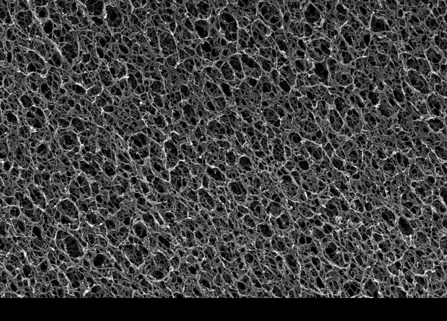
SEM image of TissueTinker Bioinks' porous microstructure. Want to read more about our bioinks? Check out our technical white paper.
Tuned to the tissue
Stiffness isn’t a detail. Increased extracellular-matrix stiffness is a hallmark of cancer: a driver of metastasis and a regulator of cell behavior through mechano-transduction. Generic bioinks ignore it. TTB-Series inks are tuned to match the mechanical stiffness and elastic modulus of the tumor at its specific organ site. Match the mechanics, and the biology follows: cells spread, form junctions, and self-organize the way they do in vivo. We let biology take the lead. (ECM stiffness and mechanotransduction)
Works with what you already have
TissueTinker isn’t a walled garden. It’s an additive biomanufacturing system that connects to the rest of your preclinical toolkit.
- Standard-compatible: glass and plastic well plates (6, 12, 96), commercial cell lines, patient-derived cells, and stem cells.
- Complementary to other NAMs: encapsulate constructs to add stromal layers to organoids and spheroids, print linked microtissues or apply flow with microfluidic organ-on-a-chip systems, or surround patient explants with printed healthy tissue.
How it compares
Set against the tools most labs use today, the difference is context: the human matrix, the tissue-matched mechanics, and the spatial architecture that older formats lose.
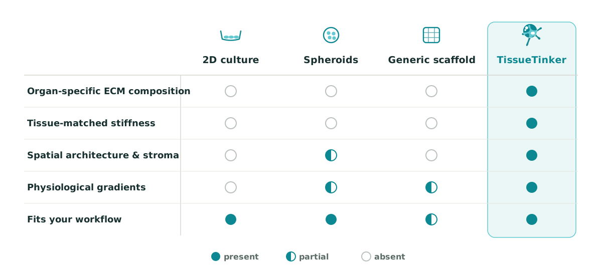
These formats aren’t rivals so much as a spectrum, and TissueTinker is designed to combine with organoids, spheroids, and organ-on-a-chip, not replace them.