
Human tumor models, printed in your lab.
Get the answers you need without leaving your building. Physiologically relevant cancer models on your benchtop, with results in weeks instead of years.
The problem: a black box
Preclinical testing runs in the dark.
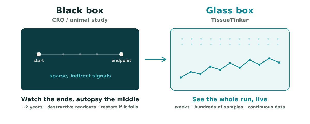
Today, answering a question about a cancer therapy usually means sending it out. A study goes to a CRO or into an animal model, and then you wait: often six months just to get on the calendar, then six to twelve more for the run. At the end there’s an endpoint, a necropsy, and the slow work of reconstructing what must have happened in between. If the data doesn’t hold, there’s no troubleshooting. You start the entire cycle again.
Two years can pass to answer a single question, and you never saw the middle.
Why it matters: the cost of the dark
It’s why most cancer drugs still fail.
That opacity has a price. Nearly 97% of oncology drug candidates fail on the way to approval. The failures trace back to preclinical models that never reflected human biology: animal studies, flat 2D culture, and generic 3D scaffolds that can’t reproduce the signaling of a real human tumor. The bottleneck was never effort. It was the model.
The shift: the door is open
The rules are changing.
Regulators agree. The FDA, EMA, and Health Canada have begun moving past animal-only testing, opening the door to new approach methodologies (NAMs) built on human biology. For the first time, the question isn’t whether to move past the old model. It’s what comes next. That’s the door TissueTinker walks through.
The solution: a glass box
TissueTinker is a glass box.
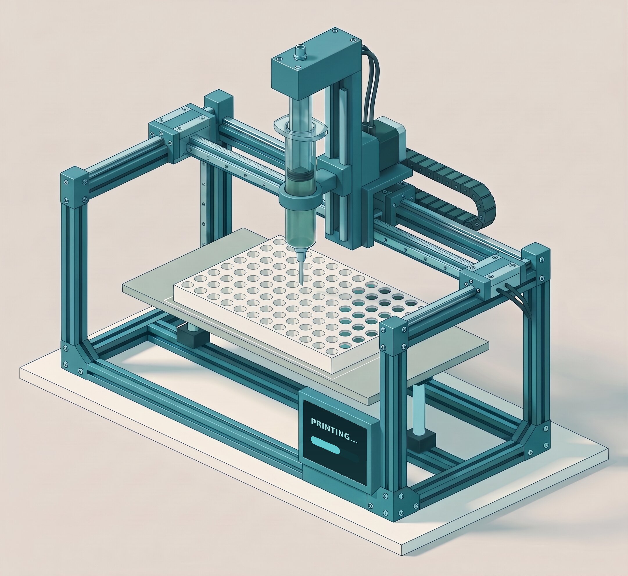
Our system prints physiologically relevant human tumor models on your own benchtop: hundreds of samples at a moment’s notice, from a printer the size of a toaster that fits inside a standard culture hood. It’s an all-in-one tool: a bioprinter that goes from box to first print in minutes, a digital library of ready-to-print tissue geometries, and our tissue-specific bioinks. Mix your cells, pick a model, print into your usual well plates, and culture, image, and assay exactly as you already do. Nothing about your workflow changes.
Instead of one endpoint two years out, you get dense, continuous data across as many conditions as you care to run. You see the middle. You stay close to your data. You iterate in weeks.
Why it’s actually better: context is the difference
The old tools flatten the biology. Ours restores it.
Animal models, 2D culture, and generic 3D scaffolds recover some of human biology, but they lose the tissue context that actually drives tumor behavior: the right matrix proteins, the right stiffness, and the conversation between a tumor and the healthy tissue and stroma around it.
TissueTinker’s bioinks are tissue-derived, not reconstituted. Each retains a real proteomic signature: collagens I, III, and IV in physiological ratios, laminin and fibronectin for adhesion signaling, and the glycosaminoglycans that shape cell-to-cell communication. Each is also tuned to match the mechanical stiffness of the tumor at its specific organ site, together restoring the biochemical conversation between cancer cells and their stroma that a one-size-fits-all matrix can’t reproduce.
And because it’s built to integrate rather than replace, the system is compatible with the rest of your toolkit: standard cell lines and patient-derived cells, and other NAMs like organoids, microfluidic organ-on-a-chip, and patient explants. It enhances the ecosystem you already work in.
The proof: what tissue-specificity actually changes
When the matrix is right, the real biology shows up.
Get the matrix right and behaviors that generic scaffolds suppress appear on their own. In our tissue-specific bioinks, and not in generic controls:
Invasion shows up.
Cancer cells switch on the markers of invasion that stay silent on synthetic scaffolds, so metastatic-like behavior becomes something you can observe.
Metabolism behaves.
Instead of runaway growth on plastic, cells grow at physiological rates and form the oxygen and nutrient gradients you need to test hypoxia-activated prodrugs and metabolic inhibitors.
Stroma does its job.
Fibroblasts remodel the matrix and lay down the dense, fibrotic barrier seen in real clinical biopsies, so you can test whether a drug actually penetrates.
Drug response looks human.
The models reproduce the reduced chemosensitivity and the heterogeneous, gradient-driven responses that separate a promising candidate from a clinical success.
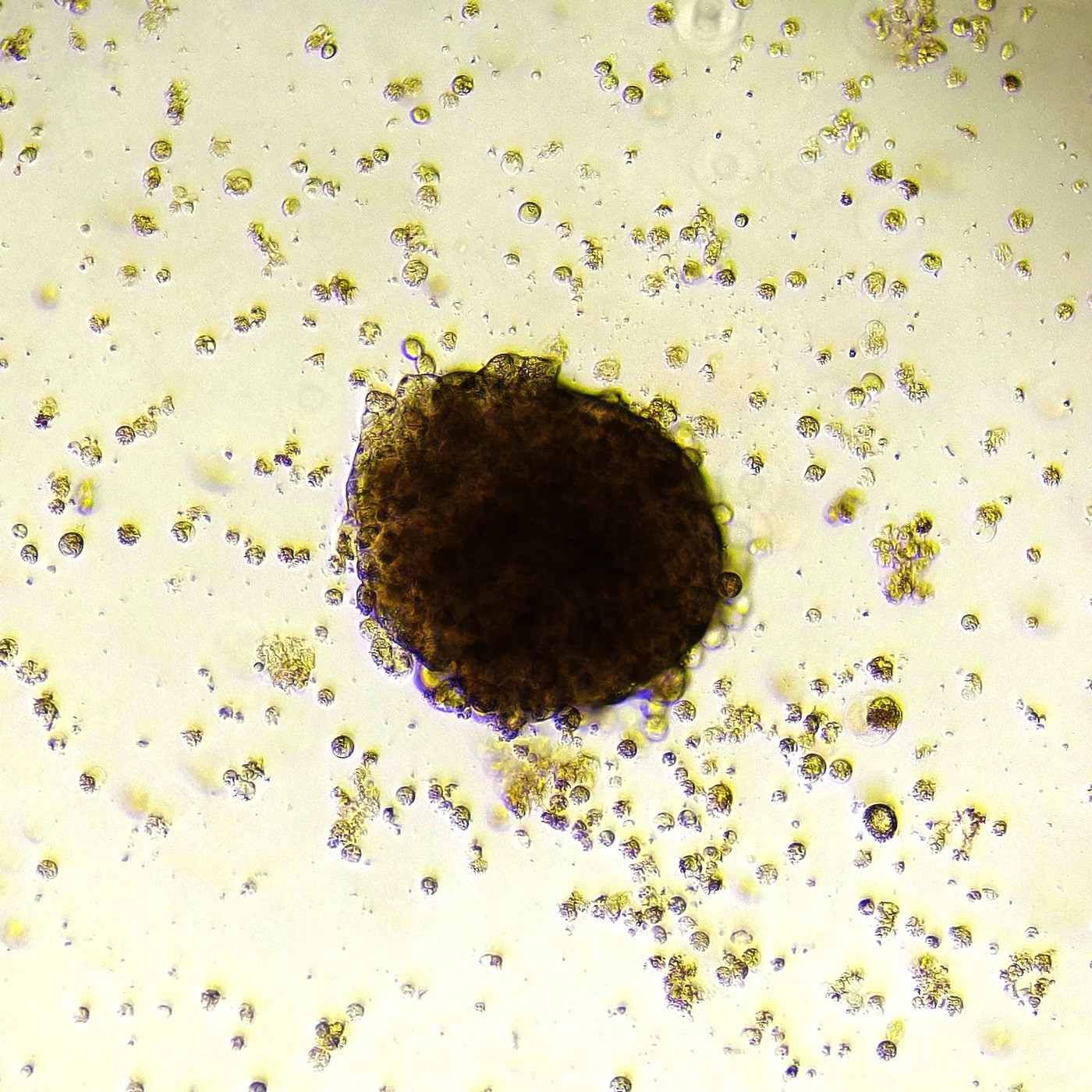
Let the matrix do the work: plated flat on our tissue-matched dECM, these colon cancer cells self-organized into 3D.
Want to hear more on the power of TissueTinker bioinks? Check out our white paper. TissueTinker grew directly out of peer-reviewed research at McGill and has been expanding it ever since.
Every tissue has a fingerprint
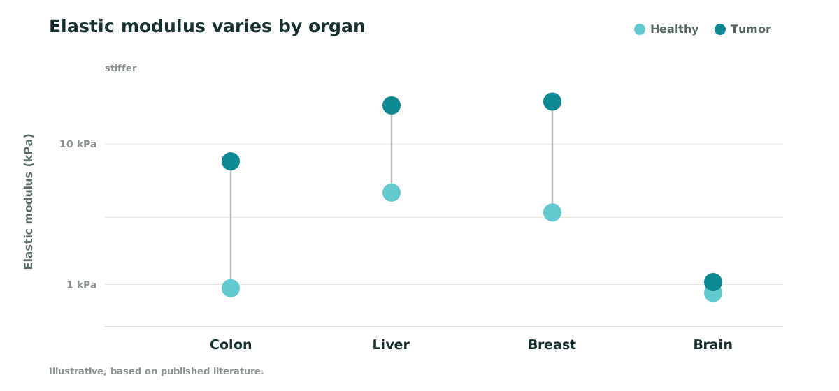
Tissue stiffness isn’t uniform.
Each organ, and each tumor, sits in its own mechanical range. TTB-Series bioinks are tuned to land in the right range for the tumor at each organ site.
Ready to see it on your bench?
We’re selecting founding early access partners. Bring your question; we’ll help you model it.
Apply for early access →Supported by










Contact
Get in touch
Partnerships, questions, or press: we’d like to hear from you.